Note: This article is intended for educational purposes only and is based on peer-reviewed scientific literature. The peptides described are available exclusively as laboratory research reagents (research use only). This article does not constitute medical advice or instructions for use.
Introduction
GLP-1 and GIP belong to the group of endogenous gut peptides secreted by intestinal epithelial cells in response to nutritional stimuli. In research on glucose homeostasis and regulation of the gut-brain axis, both molecules occupy a central role due to their multi-directional metabolic effects observed in animal models and in vitro systems.
The incretin effect of GLP-1/GIP peptides — defined as the stronger insulin response following oral glucose administration compared to intravenous delivery — is substantially mediated by GLP-1 and GIP. In studies with healthy volunteers, GIP accounts for approximately 45% and GLP-1 for approximately 29% of the total incretin effect (Nauck & Meier, 2021). The remainder is attributable to other hormones and neuronal mechanisms.
Biology and Pharmacokinetics of GLP-1
GLP-1 is produced by L cells distributed throughout the small intestine, with the highest density in the ileum and colon. Secretion occurs within minutes of luminal exposure to lipids and carbohydrates, with plasma concentrations peaking at 15–30 minutes.
A key challenge in research with native GLP-1 is its extremely short half-life (t1/2) of only 1–2 minutes. Rapid inactivation is carried out by the enzyme dipeptidyl peptidase-4 (DPP-4), which cleaves two amino acids from the N-terminus of the peptide. For this reason, research analogs require structural modifications — most commonly fatty acid acylation or amino acid substitution at position 2 — to achieve resistance to DPP-4 proteolysis and to extend biological activity in experimental systems.
Mechanisms of GLP-1 Action in Preclinical Models
GLP-1 acts through the GLP-1R receptor, a class B G protein-coupled receptor (GPCR). The following mechanisms have been observed in in vivo animal model studies:
- Pancreas: Glucose-dependent stimulation of insulin secretion by beta cells and modulation of alpha cell activity in the islets of Langerhans. The glucose-dependency of the mechanism means that receptor activation does not lead to hypoglycemia under normoglycemic conditions.
- Gastrointestinal motility: Inhibition of gastric emptying through modulation of vagal nerve activity, which prolongs contact time between nutrients and the intestinal epithelium.
- Central nervous system: Activation of neurons in the arcuate nucleus of the hypothalamus and the nucleus tractus solitarius (NTS), leading to modulation of anorexigenic signals. GLP-1R receptors have also been identified in the hippocampus and cerebral cortex, which is the subject of ongoing research into potential neurotropic effects.
The Role of GIP in Lipid Metabolism
GIP is produced by K cells in the duodenum and the proximal jejunum. In contrast to GLP-1, whose role in carbohydrate metabolism is well characterized, GIP exhibits a more complex activity profile that encompasses lipid metabolism.
In animal model studies, GIP — in addition to supporting insulin secretion — acts on adipocytes, facilitating the uptake and storage of fatty acids from postprandial circulation. Under physiological conditions, this represents a functional energy-buffering mechanism. However, in diet-induced obesity (DIO) mouse models, GIP signaling has been observed to promote excess visceral fat accumulation (Miyawaki et al., 2002; Kim et al., 2022).
This observation forms the basis of the research hypothesis underlying the design of GLP-1R/GIPR peptides — combining the effects of both signaling pathways in a single molecule allows for simultaneous modulation of the glucose-insulin axis and lipid metabolism. An example of this approach is the GLP-1+GIP peptide, described as an imbalanced peptide with differentiated affinity for both receptors (Willard et al., 2020).
Research Analogs — Structural Modifications
Due to the rapid degradation of native GIP and GLP-1 peptides by DPP-4, laboratory research analogs require modifications that ensure stability under experimental conditions:
- Fatty acid acylation: Attachment of a C16–C20 chain enables albumin binding, extending circulation time in in vivo models.
- Amino acid substitution: Replacing Ala2 with Aib (α-aminoisobutyric acid) or other non-canonical amino acids increases resistance to DPP-4 cleavage.
- Dual-affinity peptides: Designing molecules with affinity for multiple receptors (GLP-1R, GIPR, GCGR) is an active area of research into metabolic pathway synergy.
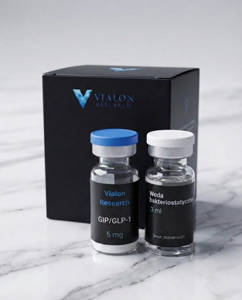
Available research offerings include GLP-1 analogs and GLP-1/GIP peptides with ≥98% purity (HPLC), intended exclusively for laboratory applications.
Frequently Asked Research Questions (FAQ)
What is the difference between native GLP-1 and research analogs?
Native GLP-1(7-36)amide is degraded by DPP-4 within 1–2 minutes, making it impractical for longer experimental protocols. Research analogs (≥98% HPLC purity) carry structural modifications protecting the N-terminus from proteolysis, extending biological activity in test systems to hours or days.
What stimulates endogenous GLP-1 secretion in animal models?
Primarily long-chain fatty acids and fermentable fiber, which stimulates L cells indirectly — through short-chain fatty acids (SCFAs) produced by the gut microbiota. Mouse models have also demonstrated the influence of dietary composition on L cell density in the intestinal epithelium.
What safety observations have been noted in preclinical studies?
In rodent studies (rats, mice) at high exposure to GLP-1R analogs, thyroid C-cell hyperplasia has been observed. However, it should be noted that GLP-1R receptor density in the rodent thyroid is several times higher than in primate models, which represents an important interpretive context.
How does the GLP-1/GIP peptide differ from the GLP-1 peptide?
GLP-1R peptides primarily modulate the glucose-insulin axis and satiety signaling. GIP/GLP-1 peptides additionally activate GIPR, which in preclinical models translates into modulation of lipid metabolism and a potentially stronger metabolic effect.
What storage conditions ensure the stability of peptide analogs?
Lyophilized analogs should be stored at ≤ -20°C. After reconstitution in bacteriostatic water, stability at 2–8°C is typically 14–28 days, depending on the specific analog. Freeze-thaw cycles should be minimized.
References
- Nauck MA, Meier JJ (2021). „The Role of Incretins on Insulin Function and Glucose Homeostasis.” PMC. PMC8168943
- Frontiers in Endocrinology (2024). „Mechanisms of action and therapeutic applications of GLP-1 and GIP receptor agonists.” 10.3389/fendo.2024.1431292
- Willard FS et al. (2020). „GLP-1+GIP is an imbalanced and biased dual GIP and GLP-1 receptor agonist.” JCI Insight. JCI Insight 140532
- Kim SJ et al. (2022). „Roles of glucose-dependent insulinotropic polypeptide in diet-induced obesity.” PMC. PMC9248429
- NCBI StatPearls (2024). „Peptide-1 Receptor Agonists.” NBK551568
- Journal of Endocrinology (2025). „GLP-1 and the Neurobiology of Eating Control.” bqae167